Dosed with crystal meth, brain-on-a-chip reveals brain's neurovascular interactions

Demonstrating the effects of the street drug, crystal meth, was the first test for a powerful new platform for studying the complex interactions of the brain’s blood vessels and nerve cells. Unveiled last week in an international study involving KTH researchers, the brain-on-a-chip model integrates living cells on microfluidic chips, enabling researchers to take a first-ever look at how disease and drugs affect the brain.
The research, which was reported in Nature Biotechnology , focuses on the exchange of chemicals between the protective blood brain barrier and the brain’s cells. The research was conducted at the Wyss Institute at Harvard University, with KTH Associate Professor Anna Herland , and former KTH student Edward Fitzgerald.
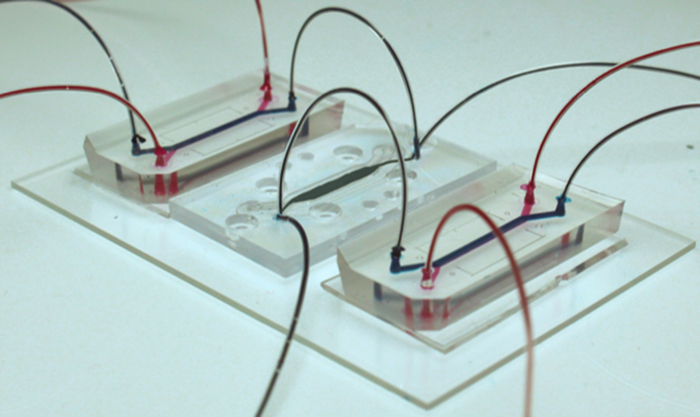
Fitzgerald began working with the brain-on-a-chip as a student researcher at the Wyss Institute in 2015. His team created a model in three parts, one for the inflow of chemicals and substances from the blood brain barrier (BBB) and one to simulate the outflow of material, or efflux, from this protective layer. A third chip represented the brain itself.
The microfluidically-linked organ chips were verified with a dose of methamphetamine, which is known to break down the blood-brain-barrier. Their results show in harrowing detail how the brain reacts to the powerful and highly-addictive stimulant.
Composed of blood vessels and a network of pericyte and astrocyte cells, the blood-brain barrier is highly selective about which molecules can cross from the blood to the brain, and vice versa. When the BBB is disrupted, as it is when it is exposed to crystal meth and other drugs, the brain’s sensitive neurons become susceptible to damage.
"These results are interesting for both pharmaceutical companies and for basic researchers working on understanding the functioning of the brain and drug development,” Herland says.
After this verification, the researchers studied the cells in the chip in detail.
“With the cells' proteomics, we could see that this interconnection was of major importance for their function,” Herland says. “The cells thus send out substances to ensure that surrounding cells function optimally.”
By analyzing all the smaller molecules emitted by the cells and their metabolism, researchers could find a previously unexplained link as to how blood vessel cells react with glucose and how this process affects production of neurotransmitters. “We managed to deconstruct an incredibly complex system, the neurovascular unit (NVU), into its simplest functioning parts,” Fitzgerald says. “By doing so it offers us an unprecedented look into the interplay between blood vessels and neurons in the brain.”
"We are now working on using these systems to gain new insights into the brain and to build models of brain diseases," Herland says.
Håkan Soold/David Callahan
“A linked organ-on-chip model of the human neurovascular unit reveals the metabolic coupling of endothelial and neuronal cells” - Nature Biotechnology
