New X-ray technology reduces radiation by 40 per cent
Greatly reduced radiation doses for both patients and doctors, less unhealthy contrast fluids and images with considerably better detail than those from conventional X-ray images. These are a few of the advantages of the technology that a KTH researcher and a number of degree project workers have developed.

The research project is called miniXpose and aims to improve the picture quality in fluoroscopy, i.e., ERCP-examinations and angiography (PCI) among other things. The tests show good results. The X-ray images speak for themselves when it comes to image quality as the increasing level of detail is impressive. It has been possible to reduce the radiation dose by 40 per cent in dental radiography in panoramic format, which the degree project worker Lahdo Aksoy discovered.
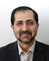
“For each coronary artery operation, the surgeon is exposed to radiation equivalent to three dental radiographs. A normally year consists of 300 operations, this corresponds to 900 dental X-rays,” says Hamed Mohammed, researcher at the School of Technology and Health at KTH and one of those involved in the research.
The degree project worker Jonatan Rogby Lindberg, is adapting the technology so that it can be used in hospitals, adds:
“The contrast fluid is harmful to the kidneys. This is important since many older patients undergo surgery and their kidneys may be a bit ”tired”,” says Jonatan Rogby Lindberg.
He adds that the technology he and the others are working on means that the amount of contrast fluid can be reduced. But there are further advantages.

“Our technique can make diagnoses more reliable, and also contribute to faster operations,” says Jonatan Rogby Lindberg.
Hamed Mohammed agrees and adds that a less anxious surgeon - because of lower radiation doses and clearer pictures - in all likelihood will do a better job.
“In addition to a better work environment due to reduced radiation doses, the protective clothing made of lead can be made lighter since less lead is required to screen the radiation. This means a more ergonomic work environment with a lower risk of occupational injuries,” says Hamed Mohammed.
The equipment itself is adaptive, i.e. it is automatic and does not require the staff to make a lot of adjustments to the controls. It deals with moving images in real time but also works equally well on single still images. There are plans to adapt the technology to other medical imaging systems, such as computer tomography (CT), in the future.
“Overall, the technology can contribute to a number of cost savings for clinics and hospitals,” says Jonatan Rogby Lindberg.
It will not be long until the technology will be available.
“The technology should be able to reach the market within 1-2 years depending on what future funding will be like,” says Jonatan Rogby Lindberg.
For more information, contact Hamed Mohammed on 08-790 48 55 / hamed@sth.kth.se or Jonatan Rogby Lindberg on 072-30 30 114.
Peter Larsson