New hope for people with a fatal diagnosis
Every year, 10 Swedish children are diagnosed with Duchenne muscular dystrophy, a fatal muscle disorder. Each year 1,000 children in Sweden are inflicted by blood cancer. Now, scientists from KTH have developed a technology which in the future will lead to patients who suffer from these two diseases having both a longer and a better life.

Today's microscopes are much better and take far more pictures than before, and the photographs can be stored digitally. More picture information provides for more complex analyses and more potential user areas. But few research groups have the time nor the resources to go through all of the material manually.
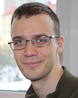
Klas Magnusson, a researcher and doctoral student at the Department of Signal Processing at KTH, has been working with colleagues at Helen Blau's Biology Lab at Stanford, in an attempt to develop a program that automates the analysis of the photographs.
"A type of analysis we have done takes one working day when automated and when using the program, but would take 10 days if we had to do it by hand, completely manually. In addition, it provides significantly more information than if we had analysed the cells manually," says Klas Magnusson.
His research project is about monitoring muscle stem cells to cause them to proliferate in a culture so that you can eventually transplant them into patients who have the deadly and progressive myopathy Duchenne muscular dystrophy.
"The technology - with some modifications - could also be useful for blood cell research and bone marrow transplantation for patients with leukaemia," says Klas Magnusson.
To get there, it is therefore necessary to know how to cultivate the cells and to be aware of how they behave when they multiply. In the current situation, often the only solution is to do the job manually. Therefore there is a substantial demand among cell biologists to be able to conduct automated analyses. Only then can we use image sequences taken with a microscope at full scale.
The automated process of analysis is based on computer vision which combines signal and image processing with programming. The first stage of the process involves image pre-processing: it is stabilized, you remove the areas of interest and then remove the background. After that the cell areas are segmented and the image is divided into background and foreground. And then you have to match the possible cells, determine which cell has travelled and where and which cells have divided in order to finally get a family tree of cells.
"In the long run what we hope to achieve is a system for the automated analysis of cells in microscope images that can be used by cell biologists everywhere, for research on a wide range of different diseases," says Klas Magnusson.
He adds that he hopes for clinical trials with the transplantation of muscle stem cells in the foreseeable future.
"There are many patients who want to participate in clinical trials, this is because Duchenne muscular dystrophy is fatal," says Klas Magnusson.
The research project is a collaboration between KTH and Stanford.
For more information, contact Klas Magnusson at 073 - 037 20 55 or klasma@kth.se.
Peter Larsson

