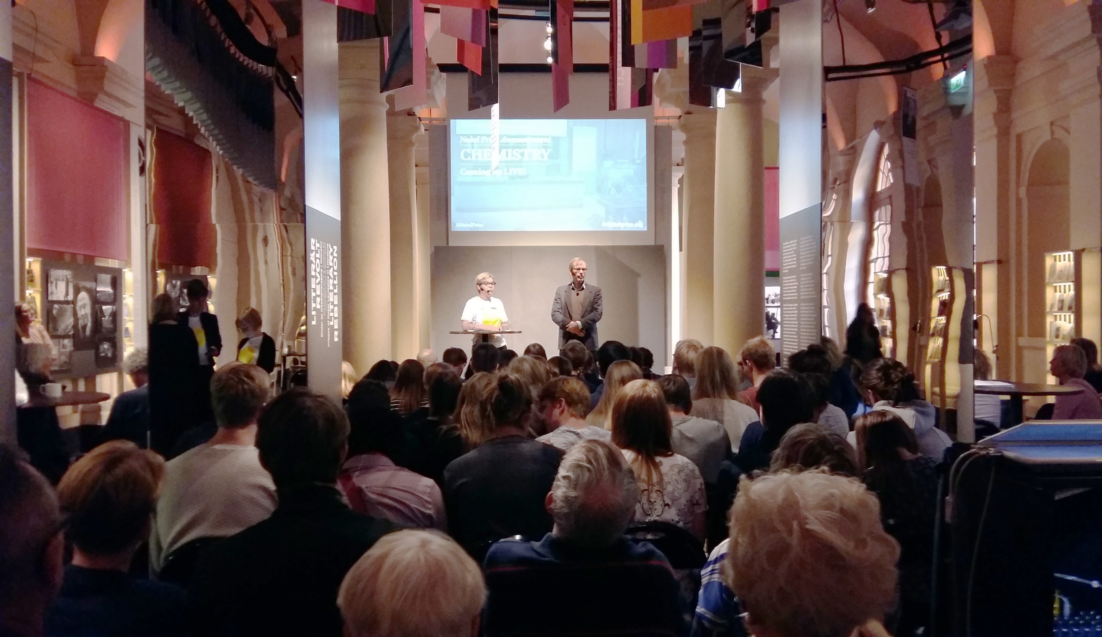
The dean explained the Nobel Prize to high school students
Hans Hebert commented this year’s Nobel Prize in chemistry and answered questions from high school students at the Nobel Museum when the Nobel laureates of 2017 were announced.

It’s lunchtime on October 4 and those who are to become Nobel Prize laureates in chemistry will soon be announced by the Royal Academy of Sciences (KVA). At the Nobel Museum in Old town, high school students from the two schools Åva gymnasium and Norra Real are seated between the pillars to watch the press conference on a screen.
Before them also stands a so far unknown expert. He has had to get into the museum incognito in order not to reveal the identity of the laureates, and not even moderator Åsa Husberg, Education officer at the Nobel Museum, knows who he is yet.
The press conference goes on air and KVA’s Sedretary General Göran K. Hansson declares that the prize is awarded to Jacques Dubochet, Joachim Frank and Richard Henderson "for developing cryo-electron microscopy for the high-resolution structure determination of biomolecules in solution". The researchers have moved biochemistry into a new era with their method, which both simplifies and improves the imaging of biomolecules, and soon we may have detailed images of life’s complex machineries in atomic resolution, according to KVA.
– Henderson’s work started in 1975 and it has taken him 40 years to get this far. It’s common that the discovery is made way back in time and then is refined, says the mysterious expert.
The man on the Nobel Museum stage makes himself known as Hans Hebert, dean of STH and professor in Structural Biotechnology. He knows the laureates from having done research in the same field since the 1970’s. Thus, he has been invited to answer the questions of the high school students and explain the laureates’ contribution to research.
– A lot of biological macromolecules such as proteins seem to function in similar ways and there is a connection between structure and function. The more we know about the structure, the more we can say about their function. There are alternative methods, but this one is more direct. You take a solution of the protein and freeze it extremely quickly as a thin, thin film. Lots of technical progress has been made and it new results are obtained at an increasingly faster rate, says Hans Hebert.
Biomolecules, the molecules of life, are molecules with biological roles, e.g. macromolecules such as proteins and carbohydrates. Cryo-electron microscopy makes it possible to see how molecules are structured on an atomic level and how e.g. proteins move, which has great importance for understanding the chemistry behind life, diseases and thereby also the development of drugs.
–Today, we can see distances between atoms of 0.15 nm, ten years ago you were satisfied with seeing 1 nm. It can concern a particular disease where a protein is failing and in order to be able to develop a drug, you need to know what the protein looks like, says Hans Hebert.
STH’s dean, who originally is a physicist, explains that research is often done in a border country and that it sometimes can be hard to decide whether a Nobel Prize should be handed out in chemistry or in medicine and to determine who should actually receive the award. A limit is set at three persons per prize.
– Richard Henderson’s lab has generated 15 Nobel Prizes. The answer usually is that they have such a good lunchroom, where they can sit and chat about research, says Hans Hebert.
How are these advances applied in everyday life? a student asks.
– Major pharmaceutical companies have got their eyes on the technique and buy their own microscopes. The more we understand about how membrane protein transport in and out of cells works and about receptors, the better we can attack diseases, says Hans Hebert.
Cryo-electron microscopy is a field that is on the rise and since 2013, when a new kind of detector was developed, new structures of biomolecules are being obtained faster. For instance, complexes for antibiotic resistance and the zika virus have been identified, and Hans Hebert’s team at STH has been involved in identifying the structure of the protein behind Huntington’s disease.
The best microscopes cost between 30 and 40 million SEK and are primarily found at national research centers, available to external users.
– New structures arrive every week and the technique is developed faster all the time. The structures are stored in a database which is open for anyone to access, says Hans Hebert.
Sabina Fabrizi