
Test shows bone fracture glue provides stronger fixation than dental filler
Acute bone fractures may soon be treated with an adhesive patch inspired by dental reconstruction techniques. Researchers at KTH report a new method which they say offers unprecedented bonding strength and a solution to the incredibly difficult problem of setting an adhesive in the wet environment inside the body.
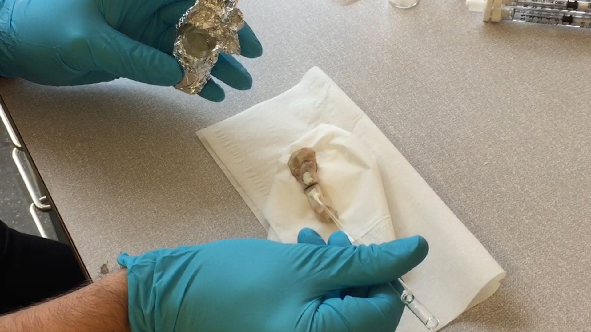
A team comprised of researchers from KTH and Karolinska Institutet report that they have successfully tested a bone adhesive on rats which combines the rigidity and load-bearing properties of dental resin composites with the bonding strength of self-etching primer – two of a dentist’s standard materials for fixing damaged teeth.
And anyone who has seen their dentist use a violet light when completing a tooth repair will recognize the technique the researchers used to polymerize, or harden, the composite material. The chemistry is called thiol–ene coupling (TEC), which ensures a bonding reaction in the presence of water.
The results offer a possible bone-setting alternative to plates and other hardware, the researchers report in the journal Advanced Functional Materials.
Michael Malkoch, Professor of Fiber and Polymer Technology at KTH, says that the new adhesive patch is biocompatible and provides bonding strength that exceeds commercially available dental bonding adhesive by 55 percent.
The technique also provides the best yet solution to the difficult challenge of fixing a patch to a wet surface without it migrating from the injury site, he says.
"We have finally managed to identify a surgically-realizable adhesive to fix bone fractures,” Malkoch says. “The chemistry, materials and methodology we used result in extraordinary adhesion and fixation to the wet bone, which in most cases is incredibly difficult.”
The patch is comprised of multiple layers: an acidic self-etching primer is laid directly on the bone’s surface to expose the collagen fibers of the bone, and to enable entanglement of the surface with the adhesive. A layer of fiber is placed on the area to be treated and then another layer of adhesive is placed on top. Finally the LED light is used for the TEC coupling. In all, the procedure takes about five minutes, he says.
With an increasing elderly population, and higher incidence of osteoporosis, doctors face a growing number of acute and complex bone injuries that require more personalized treatment. Malkoch says the technology is ready for clinical trials and he formed a startup, Biomedical Bonding AB, to drive it to market.
"We believe that the new findings will lead to a paradigm shift in fracture treatment, which in the future can phase out a large portion of today’s metal plates and screws,” he says.
The technique could offer cost savings in the form of shortened rehabilitation periods, too. Johanna von Kieseritzky and Marianne Arner, surgeons in the Department of Hand Surgery at Stockholm South General Hospital, reported that after the surgery is performed on a finger fracture, a stable fixation could allow early exercise within a day or two.
The physicians further reported that early exercise reduces the risk of fusing between the soft tissue and bone, as well as the risk of stiffness and permanent disability in the hand.
The research was funded by the Knut and Alice Wallenberg Foundation, Vinnova, Marie Skłodowska-Curie actions, and the Stockholm county government.