Ultrasound Elastography of Female Pelvic Floor Muscles: Hyperelasticity as a Biomechanical Marker of Birth Injury
This project contributes to a future where healthcare is more equal by advancing diagnostic tools tailored to women’s health—addressing childbirth-related pelvic floor muscle injuries with cutting-edge ultrasound technology. By improving detection and personalised care, the project not only aims to improve women’s health outcomes and reduce long-term suffering, but also supports a more inclusive, sustainable healthcare system.
Project description
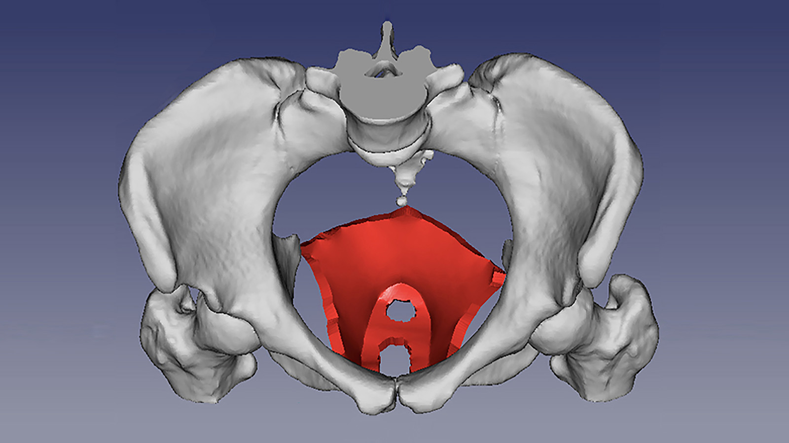
One in three middle-aged women suffers from pelvic floor dysfunction, largely due to pelvic floor injuries sustained while giving birth. This project aims to develop advanced ultrasound elastography techniques—shear wave elastography (SWE)—to assess the hyperelastic behaviour of the levator ani muscle (LAM) for enhanced prevention and assessment of childbirth-related LAM injuries.
Project goals
By developing ultrasound-based imaging tools that are accurate, affordable, and easy to implement across diverse clinical environments, the project aims to support equitable outcomes for women in a broad range of clinical and demographic contexts.
The project addresses technology development for a historically under-researched aspect of women’s health. This has the potential to result in greater gender equity in healthcare and reduced long-term complications due to birth-related LAM injuries.
Researcher
Matilda Larsson, Professor of Medical Image Processing at KTH since 2022, holds a PhD in Technology and Health from KTH (2010). Her research focuses on the development and validation of medical imaging technologies for improved diagnostic and treatment strategies in healthcare. She specialises in quantitative imaging methods in obstetrics and gynecology as well as cardiovascular applications such as mechanical characterization of soft tissue.