Imaging Surface Reactions
Catalytic surfaces can be influenced by the reactant gases, causing them to change their structures and properites. This is the origin of the 'pressure gap' between between ultrahigh vacuum surface science and applications of catalysis in industry.
We have been awarded a grant by the Swedish Foundation for Strategic Research (SSF) to develop a new instrument to study reactions on surfaces using molecular beams and velocity map imaging (VMI). The novel feature of the set-up is that it will allow the kinetics and dynamics of reactions to be studied at higher pressure than is currently possible, helping to close the pressure gap. The instrument will include a high pressure Scanning Tunelling Microscope (STM) to characterize the surface structure, helping to directly link observed changes in structre and reactivity.
News and photos of some of the big (and small) moments of progress from the lab are shown below:
Our first reactive scattering paper is accepted in Faraday Discussions: https://doi.org/10.1039/D3FD00158J We used NAP-VMI to measure CO scattering on Pd(110) and oxidation to CO2 on different O/Pd(110) oxide structures. At higher oxygen pressures, the CO oxidation reaction stops beacuse the CO liftime on the surface becomes too short.
First paper: the final version of the NAP-VMI paper is published in J. Chem. Phys. availble here https://doi.org/10.1063/5.0098495
First manuscript: read all about the new instrument here arXiv:2203.13918 where we show how Near-Ambient Pressure VMI works by scattering N2 from Pd(110) at ultrahigh vaccum and 0.001 mbar.
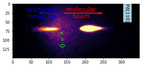
Surface scattering: we fired a molecular beam of N2 at a Pd(110) surface and detected the scattered molecules using the NAP-VMI. Molecules moving towards the surface are found to the right of the small blue cross, those on the way back to the left. The molecules make bright spots when they hit the detector. The further from the blue cross the faster the molecules were travelling.
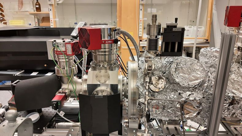
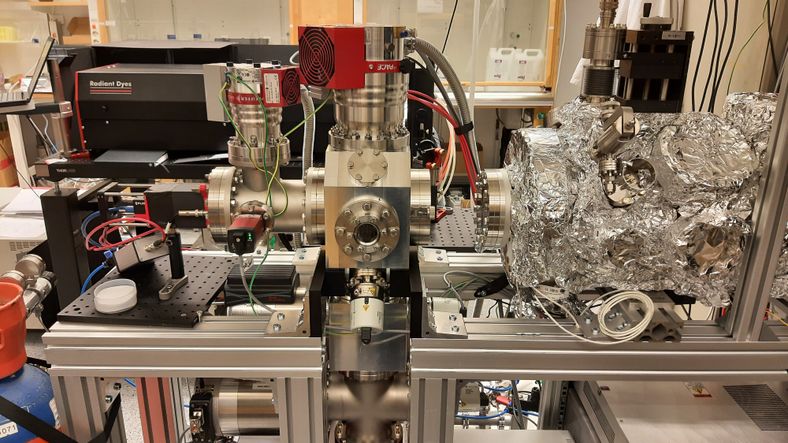
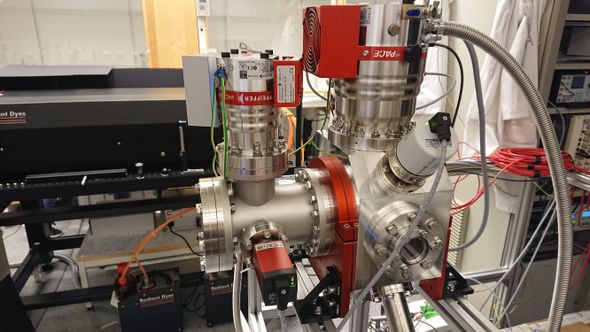
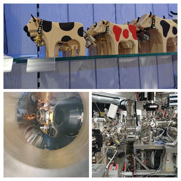
All joined together: the sample preparation chamber is connected to the rest of the machine
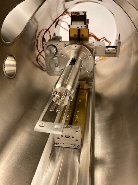
Inside, the sample holder is mounted on a UHV compatible translation statge to move between cleaning, Auger sectroscopy and scattering regions.
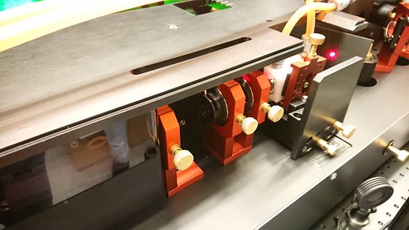
Testing the ion optics and detector.
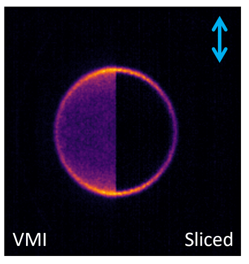
N_2^+ ions formed by resonant ionization following photodissociation of N_2O at 203 nm. The size of the ring shows how fast and in which directions the N_2 fragments were moving after dissociation. The blue arrow shows the polarization of the laser used for photodissociation and ionization.
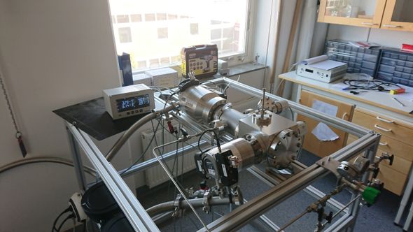
Molecular Beam and Scattering Chambers joined together
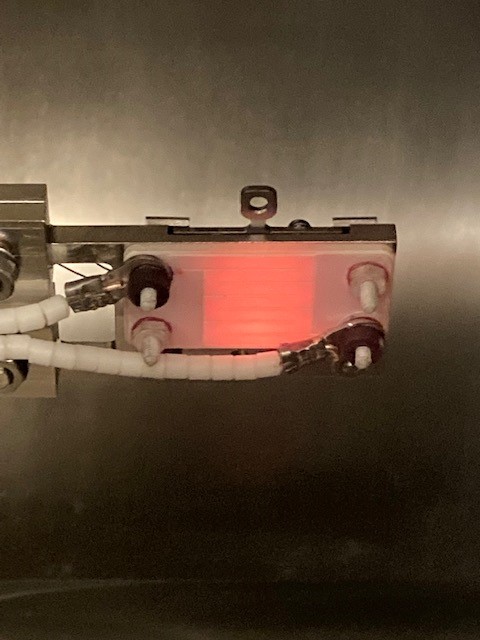
Testing the heater on the sample carrier
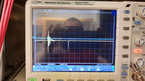
First ion signal: \({\rm N}^+_2\) ions produced by REMPI laser ionization
Ion optics mounted in the scattering chamber
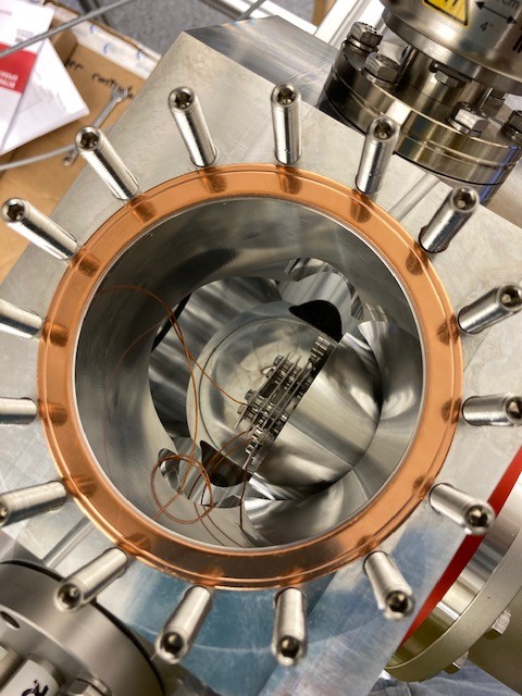
Testing the vacuum in the scattering chamber before mounting the detector. The small volume and surface area of the chamber combined with quite big pumps gives good vacuum very quickly.
First light: it works!
After a long wait, we decided to do a remote installation with video calls
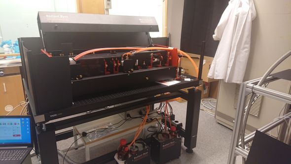
Laser system delivered, but we have to wait for travel restrictions to be eased before it can be installed.
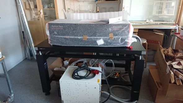
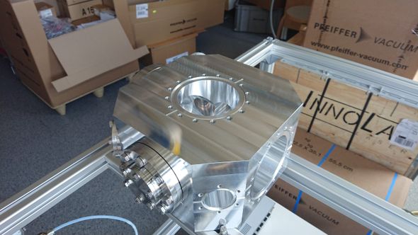
Scattering chamber delivered, this will be the heart of the new instrument
Measurement time at the PEARL beamline at the Swiss Light Source. Inspiration for building UHV instruments.
Testing the source chamber and molecular beam;