ForMAX celebrates one year – enables studies of wood-based materials from the atomic level upwards

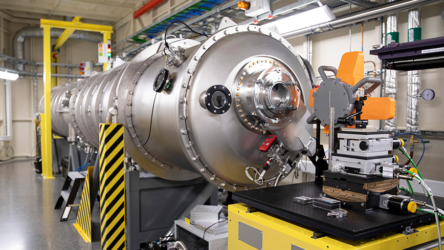
The ForMAX beamline has now been in use for a year at the MAX IV synchrotron in Lund. Researchers at KTH were the first to test the instrument, which can be used to study wood-based materials from the atomic level up to the millimetre scale.
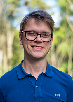
"To have one of the world's best synchrotrons in Sweden, and to have a beamline dedicated to wood-based materials is absolutely fantastic," says Tomas Rosén.
A synchrotron is a special type of cyclic particle accelerator. At the MAX IV synchrotron in Lund, there is a special instrument, a beamline, designed to study materials from the forest. The aim of the first experiment conducted by the KTH researchers in Lund, just over a year ago, was to test the capabilities of the instrument and see if it was suitable for some of their most demanding experiments.
"We are particularly interested in looking at flows of cellulose nanofibrils, and studying processes where they are assembled into super-strong materials," says Tomas Rosén, researcher at the Department of Fibre and Polymer Technology at KTH.
Down to the nanometre level

With the techniques offered at ForMAX, the researchers can see how the nanofibrils organise themselves in the flow at a nanometre level. However, in these processes, the concentration of nanofibrils is very low, around 0.2% by weight (99.8% water), which means that the background signal from the instrument has to be very low to get useful data. To the researchers' delight and partial surprise, ForMAX was able to deliver this from day one, with comparable background levels to the best beamlines they had previously used for the same type of measurements.
“In our case, we send the X-ray beam through the flow of cellulose nanofibrils and study the resulting scattering pattern. From this we can obtain statistics on the orientation, aggregation and cross-sectional sizes of the fibrils; information that we can then directly compare with numerical simulations to optimise our material process," says Tomas Rosén.
“By looking at X-ray scattering at larger scattering angles, it is also possible to simultaneously find out about structural changes at the molecular level, and in our case how cellulose polymers arrange themselves inside individual nanofibrils.”
A multidisciplinary instrument
What has it meant for the researchers at KTH to be able to use this instrument? According to Tomas Rosén, interest in synchrotron experiments has grown steadily in recent years, making it increasingly difficult to obtain beam time. Researchers have previously had to travel with all their equipment to facilities in the USA, Japan, Switzerland and Germany to make their measurements. The fact that research on these instruments is so multidisciplinary means that you cannot expect the staff on the beamlines of synchrotrons to know much about cellulose, for example. This is exactly what makes ForMAX so special; that it is dedicated and designed specifically for materials found in the forest.
“It is great to see how many colleagues find new exciting ideas to investigate with synchrotron light and the ideas are also starting to reach the industry, which is extra fun to see! In my role, I now continue to spread knowledge about what can be done with these instruments and try to help our partners as much as I can with designing experiments and analysing data.”
Text: Jon Lindhe

