MTH microCT - miniPET
Technology description
The microCT main components are a 120 mm x 120 mm flat-panel detector (Hamamatsu CMOS flat panel C7942CA-22) with a pixel size of 50 μm and a maximum frame rate of 2 Hz at full resolution, an x-ray tube (Hamamatsu microfocus X-ray source 90 kV, pulsed MX-L10951-04 ) that can be operated in pulse mode with a focal spot size of 15-80 μm depending on the actual power used.
It is possible to change the source-to-object distance in order to image small animals (or parts of them) with different magnification factors.
The spatial resolution for 2D imaging has been measured to be 7 lp/mm, while for 3D imaging the resolution strongly depends on the particular imaging task.
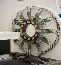
The miniPET ring consists of 12 detector modules made of LYSO scintillator pixelated crystals (1,27 x 1,27 x 12 mm3) in a 35x35-pixel matrix (pitch 1,347 mm). The crystals are optically coupled to SiPM:s (ST-microelectronics).
Preliminary results show a spatial resolution at ¼ CFOV of 1,24 mm (tangential), 1,15 mm (radial) and 1,23 mm (axial), a peak absolute sensitivity of 1,36% (0,7% total absolute sensitivity) and a NEC-peak for the rat phantom of 24 kcps (at 43 MBq) and of 91 kcps (at 44 MBq) for the mouse phantom.
In order to maintain full control over the system, the acquisition and the reconstruction software for both the CT and the PET have been written by us and are going to be continuously upgraded. Hardware upgrade will be done depending on fundings.
Clinical impact
The microCT/miniPET can image give both morphological (CT) and functional (PET) images of small animals. It can also be used for non-destructive inspections of ex-vivo samples (e.g. arterial plaques). The CT and PET can also be used separately.
The main advantage of this scanner compared to other commercial solutions is that we can adapt it to fulfill the needs of the researchers that intend to use it (and we are really looking forward to hear from researchers that have particular requests!).
The PET ring is MRI-compatible and could in principle be used for PET-MRI studies.
Application specialist
Contact person
For more information please contact the Jonassons Medical Imaging Centrum