
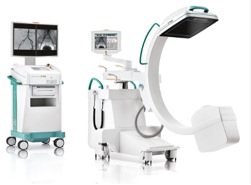
Mobile C-arm. Ziehm Vision RFD.
Technology description
This system combines the full flexibility of a mobile C-arm with fixed room imaging capabilities. It offers increased clinical confidence thanks to distortion-free image and expanded anatomical visualization from the latest 30 cm x 30 cm flat-panel technology. The square shape of the flat-panel increases the field of view.
The C-arm utilizes to high-power pulsed fluoroscopy monoblock generator for optimal anatomic penetration with minimal dose of ionizing radiation. The system is equipped with a unique liquid cooling system for extended performance in demanding clinical procedures.
The construction simplifies patient positioning, handling and dose control. Object Detected Dose Control (ODDC) technology creates a matrix over the entire scan field and uses 256 measurement cells to scan the region of interest in real time. All settings, including the dose level and noise filters, are automatically adapted to the patient’s position and also automatically adjusted to eliminate motion artifacts or to reduce the image distortion.
Technical features:
- More than 16,000 shades of gray for greater contrast
- Expanded dynamic range: 84 dB
- Improved vessel resolution and soft tissue definition
- 1.5k x 1.5k image matrix
- Compact and powerful monoblock generator with up to 20kW
- Rotating anode
- 4 ms – 50 ms high frequency pulse width
- Pulse rate up to 25 frames per second
- Small footprint (0.8 m2)
- Easy-drive system for easy maneuverability
- Single lever steering and braking
- Deeper C-arm opening
- 165° rotation with counterbalanced C-arm
Clinical impact
The mobile C-arm, Ziehm Vision RFD, offers great flexibility and is exclusively designed for demanding procedures in:
- Cardiovascular Surgery
- Endovascular Surgery
- Interventional Cardiology
- Interventional Radiology
- Neuro and Spine Surgery
- Coronary Angioplasty
Short, powerful pulses up to 20kW offer superior visualization of moving objects as needed in cardiac procedures. In combination with the high dynamic range and high resolution of the flat-panel detector, the equipment provides visualization of even the smallest vessels. Specially designed vascular software helps to provide good image quality in all vascular procedures.
Contact person
For more information please contact the Jonassons Medical Imaging Centrum