
Photoacoustic Imaging Facility
Technology description
Photoacoustic imaging is a novel in vivo hybrid imaging modality that combines the sensitivity and contrast of optical imaging with the depth and resolution of ultrasound. When pulsed laser light illuminates tissue, the optical absorbers, i.e. hemoglobin or contrast agents, undergo thermoelastic expansion, generating an acoustic pressure wave, which is detected with an ultrasound transducer.
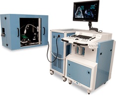
The photoacoustic imaging system consists of the high-frequency ultrasound digital imaging platform Vevo 2100, the laser control system Vevo LAZR and an imaging station with an injection mount covered by a laser protection hood.
This digital platform delivers outstanding performance in a wide range of applications including, but not limited to, cardiovascular, cancer and inflammation models. The new MicroScan transducers provide increased frame rates and resolution, superb contrast, and a wide field of view. The photoacoustic imaging system is an intuitive, user-friendly system that allows a high degree of flexibility and customization for every application. It is non-invasive and fast, providing extremely high throughput when needed.
New solid-state transducers
- Superior resolution and image uniformity through the entire field of view
- 30 micron resolution
- Frame rates in 2D up to 740 fps (for a 4x4 mm field of view)
- Wider field of view
Expandable Imaging and Processing Options
- Color and Power Doppler Modes for blood flow quantification & anatomical identification
- M-Mode single line acquisition allowing high-temporal resolution for LV functional analysis
- Anatomical M-Mode for adjustable anatomical orientation in reconstructed M-Mode imaging
- 3D-Mode Imaging & Volume Analysis
- Nonlinear Contrast Imaging
- VevoStrain™ Analysis software for cardiac research
- Advanced measurements & quantification
- Open architecture data management, flexible image output options
- Mobile - true plug & play for any lab
The ultrasound imaging provides a high-resolution frame-of-reference for identifying anatomy, while the photoacoustic imaging enables functional measurements such as oxygen saturation, total hemoglobin and the microdistribution of biomarkers. All tissue can be imaged in real time and in vivo down to 2 cm in depth with a resolution down to 45 μm.
In addition, the optical imaging capabilities of photoacoutics can be used for contrast applications with dyes as well as molecular imaging applications with nanoparticles. Both ultrasound and optical photoacoustic signals are co-registered, allowing users to track the location of multiple molecular signals detected with dyes or nanoparticles back to the anatomy.
Advantages of Photoacoustic Imaging
- Optical contrast for blood and molecular imaging
- High-resolution at depth
- Real-time
- Non-invasive
- Anatomical, functional and molecular data
Clinical impact
The equipment enables the researcher to obtain in vivo anatomical, functional, physiological and molecular data simultaneously, in real-time and with a resolution down to 30 µm.
Photoacoustics is a powerful modality and add-on to the high-frequency ultrasound technology. It works in combination with ultrasound – in real-time and non-invasively – to go beyond basic imaging. Structural information from the traditional B-mode ultrasound imaging is combined with spatial distribution of blood flow and characterization of the blood flow in the PA-Mode. Areas of application includes:
- hypoxic state of the tumor microenvironment to measure hypoxia and angiogenesis in cancer imaging
- vascular networks
- obstetrics, including fetal and maternal physiology
- stroke/ischemia and functional brain imaging studies
Contact person
For more information please contact the Jonassons Medical Imaging Centrum