
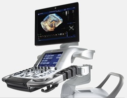
Vivid E9 Ultrasound system
Technology description
This cardiovascular ultrasound imaging system is specifically designed for 4D imaging. The system is equipped with advanced quantification tools such as 4D Stress and 4D Auto LV quantification.
The core of the system is GE’s beam-forming technology, the Accelerated Volume Architecture, that in combination with both coherent and harmonic image processing provides computational power, ease of imaging and workflow flexibility. It is equipped with 5 new imaging transducers—two designed with XDclear transducer technology— for adult, pediatric, vascular, and abdominal applications. The XDclear technology introduces enhancements in both 2D and color-flow image quality. Moreover it enables the visualization of pulmonary vein inflow. For example, the 9L-D transducer delivers enhanced signal-to-noise ratio, reducing noise in the vessels. The 8C transducer is also well suited for vascular imaging. It supports carotid and vertebrae imaging where access with conventional wider aperture linear transducers may be limited by anatomical barriers.
Quantification tools include:
- Automated Function Imaging (AFI) to assess and quantify left ventricular wall motion at rest. It calculates a large set of parameters to describe the function of the left ventricular walls. AFI specifically calculates peak systolic longitudinal strain (both segmental and global) and presents the results as parametric images.
- 4D Strain to calculate both global and regional strain values based on a spatial speckle tracking algorithm. The end result is presented in a Strain Bull’s Eye plot accompanied by time-strain curves and cut planes for enhanced visual tracking assessment.
Other tools include:
- 4D Auto Left Ventricle Quantification (LVQ)
- 4D Left Ventricle (LV) Mass
- Mitral Valve (MV) Assessment
- 4D Views to view images such as 4-, 2- chamber APLAX, mitral valve, septum and aortic valve from one full volume acquired data
- Scan Assist to customize the system for the department protocols
Clinical impact
Visualization of the results and translation of the knowledge from sonographers to other clinical experts is another critical aspect that is taken care within Vivid E9 platform. GE has standardized Depth Illumination on this platform to enhance communication to non-echo experts. The new depth color map helps visualization when the object is illuminated by an imaginary light source, casting a shadow enhancing depth/distance perception. This can be very helpful for the surgeon in the OR or the interventionalist in the cath lab to ensure smooth procedure in a crowded and stressful environment. Depth perception is enhanced by a visualization technique called Polar Vision, which combines polarized stereo with depth rendering on a dedicated 3D monitor.