
Division of Biomedical Imaging
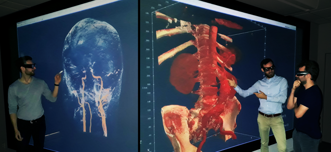
Biomedical imaging comprises a whole pipeline consisting of data acquisition, image reconstruction, image enhancement, image analysis, image-based simulation and visualization for solving a problem within clinical diagnostics or medical research. At the Division for Biomedical imaging we deal with all of these steps in our research and teaching.
Highlights
Overview

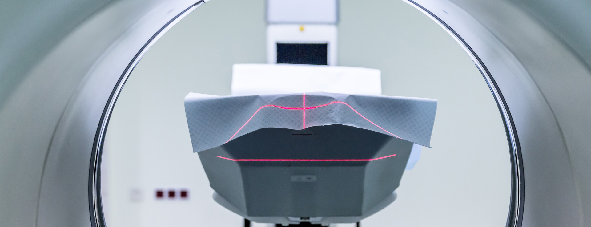
Acquisition of data and image reconstruction algorithms are central in our work with Positron Emission Tomography integrated with Computed Tomography (PET/CT), where we have designed KTH’s own miniPET/microCT system, to be used in animal experiments. New ultrasound-based techniques for detection of cardiovascular disease are being developed and tested in phantoms, animals and humans. Other research aims at developing new contrast agents useful for several imaging modalities.
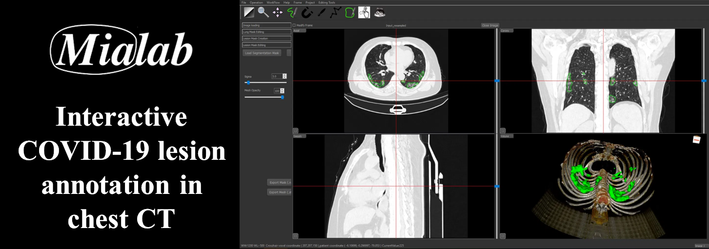
We also work with post-processing of medical image data, often using techniques from machine learning (such as deep learning), to derive quantitative measures (imaging biomarkers) for diseases such as dementia or cancer, mostly from Magnetic Resonance Imaging (MRI) data. Image-based simulation can be used for estimating mechanical properties such as bone strength from high-resolution in vivo images. Machine learning is also applied for image enhancement in angiographic interventions.