To KTH's start page To KTH's start page

Our projects

Acoustic Response of Pickering Perfluoropentane Droplets

This project aims at understanding the acoustic response of these pickering perfluoropentane droplets. We study how the threshold for acoustic droplet vaporization changes between bulk and confined medium, investigate how various parameters influence the resonance frequency of these droplets, and develop an imaging sequence that successfully image the droplets before and after vaporization.

Characterization of the mechanical properties of the brain through magnetic resonance elastography (MRE)

This project aims at characterizing the mechanical properties of the brain through MRE in adolescents and older children and at correlating them with risk factors for developing anxiety disorders. Our long-term goal is to improve the diagnosis and treatment of anxiety disorders in the young population.

Synthetic MRI aging

One challenge in image analysis of Alzheimer’s patients is to disentangle the effects of aging from the disease itself. For this, it is necessary to use a large number of images of healthy subjects at every specific age, which is not available in the current longitudinal imaging datasets. In this project, we will develop an AI-based solution to generate magnetic resonance images (MRI) at different ages by simulating the changes in the images due to aging.

The required energy for the pharmaceutical removal in the cavitation reactor

This project focuses on the energy requirements for wastewater treatment regarding pharmaceutical removal with hydrodynamic cavitation. The main objective of the project is to identify the mechanism responsible for the pharmaceutical compound decomposition due to the collapse of cavitation bubbles.

Methods for the analysis and characterization of brain morphology from MRI images

Brain magnetic resonance imaging (MRI) is one of the most commonly used methods for investigating brain morphology, since it allows to produce detailed 3D images of its soft-tissue structures. In this project, we are working on the development of methods for the analysis and characterisation of brain morphology from brain MRI images.

Improved treatment of tumors with novel image tools

In the treatment of cancer, which is one of the greatest threats to public health, new treatments are continually being developed.Our research aims at developing tools that are needed for planning of surgery and radiotherapy and for evaluating the effect of treatment regimes as early as possible. This includes new segmentation methods based on machine learning.

Improved image tools for describing diseased and healthy blood vessels

With Computed Tomography Angiography (CTA), blood vessels such as the coronary arteries can be studied. Here we develop new methods for segmenting and measuring the lumen (blood-filled cavity) and the wall of the vessel, aiming at higher accuracy and lower radiation doses.

Patient-specific left ventricular flow simulations from transthoracic echocardiography

This project aims to develop and evaluate a pathway for patient-specific computational fluid dynamics (CFD) modeling of the left ventricle using 4D transthoracic echocardiography (TTE) as input modality.  

Shear Wave Elastography for Vascular Applications

The aim of this project is to develop and evaluate ultrasound shear wave elastography in vascular applications, both to assess arterial stiffness and to perform atherosclerotic plaque characterization.

CACTUS - Contrast Agent for CT and Ultrasound: manufacturing and characterization

Hybrid imaging is defined as a powerfull combination of two or more imaging techniques. By doing so, the most relevant properties from each modality can be merged together. For instance, the high resolution of CT can be combined with the high temporal resolution of US.

To increase further the sensitivity and specificity of hybrid imaging, an injectable microdevice supporting multimodality imaging approaches would be of great value.

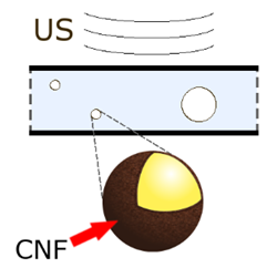
In the CACTUS project, we develop a micro/nano-construct that can support multimodal imaging. The two modalities that our device is intended for are CT and US, but other imaging techniques such as photoacoustic or optical imaging could be also supported.

CATAMARAN - Contrast Agents for Theranostics of Atherosclerosis using Microbubbles and Recent Advanced Nanotechnology

The translational significance of this study is to develop a simple combined tool for diagnostics and treatment of acute myocardial ischemia in patients with atherosclerosis or diabetes with impaired bioavailability of NO. Keeping in mind that systemic treatment with NO or NO-donor has a very narrow therapeutic window and may provoke critically low blood pressure, novel approach for NO transport, local delivery and triggered administration is of high clinical interest.

MUMI2D - Microdevice for ultrasound mediated imaging and drug delivery

The current project introduces a new class of microdevices providing integrated diagnostic and therapeutic applications, i.e., theranostics using microcapsule/microbubble based ultrasound contrast agent loaded with therapeutic compound. Application of perfluorocarbon as a core media facilitate the acoustic droplet evaporation which is transition from capsules to bubbles under specifically designed ultrasound pulse. A gas core makes microbubbles an efficient ultrasound contrast agent for imaging. Therapeutic compound might be mixed with perfluorocarbon and delivered locally following high energy ultrasound pulse and burst of the bubble.

Tendon biomechanics

The aim of this project is to develop quantitative methods to be used in studies of tendon biomechanics. The research questions stem from orthopedically based clinical needs. The purpose of the project is to evaluate the effects of treatments for ruptured tendons, but also tendon biomechanics in non-injured individuals.  

Analysis of Bone Images

The economic burden of osteoporosis-related factures on public health systems is huge, and it is actually more severe in Nordic countries. Osteoporosis is a disease that weakens the internal structure of trabecular bone. Thus, it is of utmost importance to devise quantitative assessment tools for analyzing 3D images of trabecular bone. The emergence of high resolution imaging modalities, especially Cone Beam Computed Tomography (CBCT) and High Resolution Peripheral Quantitative Computed Tomography (HR-pQCT) has opened the door to perform such analyses in vivo. We have developed in the last few years several methods for analyzing the microstructure of trabecular bone in gray-scale, including trabecular thickness quantitation, classification of trabeculae and estimation of different types of fabric tensors. The methods have already been validated by comparing them to an established reference method (micro-CT) in bone specimens. However, before these methods are used in clinical routine, it is necessary to perform an extensive validation, which is one of the objectives of this project.