Shear Wave Elastography for Vascular Applications
Description (background and methods)
For patients with carotid artery disease, current guidelines state that endarterectomy (surgical plaque removal) is highly beneficial in patients with carotid stenosis of >70% occlusion. However, recent studies demonstrate that plaque composition, rather than the degree of stenosis, is essential in emboli formation. Soft plaques, which are characterized by a thin weak fibrous cap, intraplaque hemorrhage, and large lipid core, are associated with a higher risk of rupture than stiffer, more stable plaques. In vivo mapping of plaque mechanical properties would therefore be of particular interest for improved diagnostic and treatment strategies. In addition, accurate non-invasive methods to assess early signs of cardiovascular disease, such as increased arterial stiffness, are lacking.
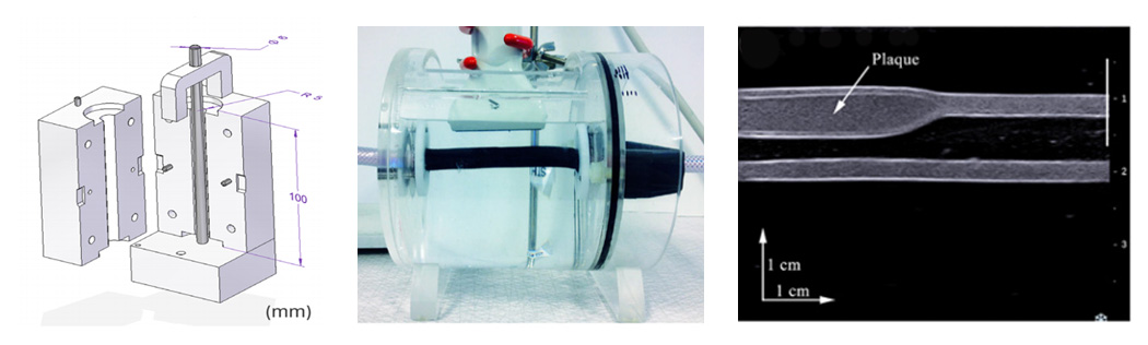
For this purpose, we have developed shear wave elastography for arterial applications. In shear wave elastography, an acoustic radiation force is generated in the tissue that in turn results in a transverse low frequency propagating shear wave. The speed of the shear wave is measured by high speed ultrasound imaging and then related to the mechanical properties of the material. The project concerns adaptation and validation of this technique to measurements in the arterial wall, since the technique has mostly been used in uniform large stationary tissues before, where less dispersion and wave reflections are present. The accuracy of the technique has been studied in comparison with mechanical testing in pressurized arterial phantoms with and without plaques with promising results. Also, the influence of a confined geometry on the estimated mechanical properties has been assessed by experiments in phantoms and by finite element method simulations, and the sensitivity of the technique has been studied in small porcine aortas used as a human carotid artery model.
In on-going studies we investigate the safety of the technique when applying it to the arterial wall, by estimating the strain and strain rate generated by the acoustic radiation force impulse. Also, wave propagation in different phantom plaque geometries when generating the acoustic radiation impulse at different spatial locations is being investigated.
(Preliminary) results and publications
More information about the results from the projects can be found in the following publications:
Peer-reviewed publications
- Maksuti E, Bini F, Fiorentini S, Blasi G, Urban MW, Marinozzi F, Larsson M. Influence of wall thickness and diameter on arterial shear wave elastography: a phantom and finite element study. Physics in Medicine Biology, 2017; 7;62(7):2694-2718.
- Widman E, Maksuti E, Amador C, Urban MW, Caidahl K, Larsson M. Shear Wave Elastography Quantifies Stiffness in Ex Vivo Porcine Artery with Stiffened Arterial Region. Ultrasound in Medicine and Biology, 2016;42(10):2423-35.
- Maksuti E, Widman E, Larsson D, Urban MW, Larsson M, Bjällmark A. Arterial Stiffness Estimation by Shear Wave Elastography: Validation in Phantoms with Mechanical Testing. Ultrasound in Medicine and Biology. 2016;42(1):308-21.
- Widman E, Maksuti E, Larsson D, Urban MW, Bjallmark A, Larsson M. Shear wave elastography plaque characterization with mechanical testing validation: a phantom study. Physics in Medicine Biology, 2015; 60, (3151-3174).
Conference proceedings
- Maksuti E, Larsson D, Urban M, Caidahl K, Larsson M, Strain and Strain Rate Generated by Shear Wave Elastography in an ex vivo Porcine Aorta, IEEE International Ultrasonics Symposium, 2017, Washington, USA.
- Larsson D, Roy J, Urban MW, Colarieti-Tosti M, Larsson M, An ex-vivo setup for characterization of atherosclerotic plaque using shear wave elastography and micro-computed tomography, IEEE International Ultrasonics Symposium, 2016, Tours, France.
- Widman E, Maksuti E, Amador Carrascal C, Urban M. W, Larsson M. Evaluating Arterial and Plaque Elasticity with Shear Wave Elastography in an ex vivo Porcine Model. IEEE International Ultrasonics Symposium 2015; Taipei, Taiwan.
- Widman E, Matsuki E, Larsson D, Caidahl K, Urban M, Bjällmark A, Larsson M, Feasibility of Shear Wave Elastography for Plaque Characterization – An experimental study using Mechanical testing. IEEE International Ultrasonics Symposium; 2014; Chicago, USA.
- Widman E, Maksuti E, Larsson M, Bjällmark A, Caidahl K, D’hooge J. Shear Wave Elastography for Characterization of Carotid Artery Plaques – A feasibility study in an experimental setup. IEEE International Ultrasonics Symposium; 2012; Dresden, Germany.
Collaborations
- Mayo Clinic College of Medicine, Rochester, MN, USA
- Matthew Urban, Associate Professor of Biomedical Engineering, Department of Radiology
- Karolinska Institutet, Stockholm, Sweden
- Kenneth Caidahl, Professor, Department of Molecular Medicine and Surgery
Thesis projects
- Assessment of Shear Wave Elastography Acoustic Output - a Simulation and Experimental Study, Cristiana Golfetto, 2016
- Accuracy Assessment of Shear Wave Elastography for Arterial Applications by Mechanical Testing, David Larsson, 2014
- Comparison of Pushing Sequences for Shear Wave Elastography, Tim Nordenfur, 2013
Contact person

Contributors