Molecular imaging of cancer
In vivo molecular imaging has potential to improve the diagnosis of different diseases by defining relevant aberrant cellular alterations and to indicate adequate treatment. In cancer diagnostics, molecular imaging can provide a global view of potentially all metastatic lesions in the body. Radionuclide molecular imaging of therapeutic targets is a non-invasive procedure that is easily repeated to follow the effects of a treatment regimen. Radiolabeling of monoclonal antibodies (mAbs) is a straightforward way to develop imaging agents, but mAbs have the inherent drawback of long residence time in circulation, which results in poor imaging contrast. Small imaging probes provide higher contrast, sensitivity, and faster analyses than full-length mAbs due to better tissue penetration, faster blood clearance, and reduced nontarget-specific accumulation in tumors due to minimal influence from the ‘enhanced permeability and retention’ effect (EPR effect).
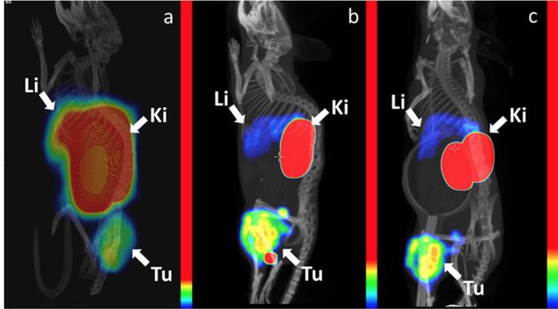
We are investigating affibody-based probes for molecular imaging of tumor-antigens, such as EGFR, HER3, CAIX and VEGFR2. The research is performed in close collaboration with the research groups of Profs Vladimir Tolmachev and Anna Orlova at Uppsala University.
Research funded by: Vinnova - Sweden's innovation agency