

Method makes it possible to single out and attack HIV virus’ most dangerous parts
A new method could make it possible to identify the most dangerous parts of the HIV virus, so they can be singled out for attack.
Researchers at KTH Royal Institute of Technology have created a method of illuminating viral molecules that blink on and off, enabling more accurate measurements of a virus’ progressive growth than currently possible. KTH researcher Ilaria Testa says the method makes it possible to track which molecules in the HIV virus are essential for growth.
“That knowledge could be put to use in finding therapies for HIV positive patients, since it can help identify which molecules to block to prevent the virus from growing,” Testa says.
The research was reported last week in the scientific journal, Nature Biotechnology. It was carried out at the Science for Life Laboratory (SciLifeLab) in Stockholm.
The key to identifying whether molecules are binding to other proteins is to measure how fast the molecules rotate. Just as larger objects usually rotate slower than the smaller ones, the more mass a molecule accumulates the slower it will rotate.
KTH Associate Professor Ilaria Testa says the method improves upon a commonly-used form of fluorescence technique which can only measure fast rotation of molecules. But such techniques are blind to the typical, slower rotation of human proteins.

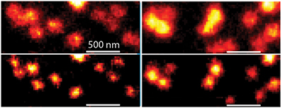
The researchers doped an HIV viral particle with specially-engineered fluorophones that switch themselves on and off so that they highlight the orientation of the molecule. With longer on and off states these fluorophones—or reversibly photoswitchable fluorescent proteins—enable the identification of slow rotation of growing molecules.
“We showed that the method can be used to identify HIV maturation state and to learn more about how viruses like HIV progress,” Testa says.
The research was financed with the support of the Swedish Foundation for Strategic Research (Project no. SAB 19-0033 and FLL15-0031), the Göran Gustafsson Foundation (GGS1709), the Marie-Curie EU project (no. 895938/STARSS), Deutsche Forschungsgemeinschaft (project no. 240245660) and SFB 1129 (project 6).
David Callahan