
Millions to KTH for research on molecular medical imaging
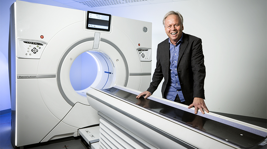
KTH Royal Institute Professor Mats Danielsson's research team has been awarded a European Research Council Advanced Grant of €3,351,875 for a five-year project that could revolutionize hospital technology for PET and SPECT, known as molecular imaging. Among other uses, the methods can detect cancer and cardiovascular diseases at an early stage.
Danielsson , Professor of Medical Imaging Physics at KTH, is one of the selected research leaders to receive one of Europe's most prestigious research grants, ERC Advanced Grants , a grant from the European Research Council.
Danielsson likens the new technology to a camera that records molecular medical images." The new technology means that the efficiency for detection of individual gamma rays can be increased by up to a million," Danielsson says.
“The time it takes to record an image of the patient can be reduced from 10 minutes to less than a second.”
Individual molecules can be tracked
Molecular imaging in the form of SPECT (Single-Photon Emission Computed Tomography) and PET (Positron Emission Tomography) is a commonly used examination in healthcare today, including for the early detection and follow-up treatment of cancer. The researchers now hope that the new technology through higher resolution will make cancer treatment more effective.
"Through this research project, the radiation dose that patients are exposed to can also be radically reduced, because the new camera is much more efficient," Danielsson says.
“Our new technology can also be used to diagnose cardiovascular diseases, for example. Unlike conventional X-ray technology, individual molecules can be tracked, and body functions, such as metabolism, can be studied almost in real time.”
The research team expects the new technology to contribute to faster development of new and more effective drugs, where SPECT and PET already play an important role.
The group has been collaborating for several years with doctors and engineers at
MedTechLabs
.
“The ERC grant supports our basic research, and we believe that the new technology can be used in healthcare within five to ten years," Danielsson says.
Out of over 1 800 applications for the ERC Advanced Grant, 255 projects were selected.
"We are delighted to have succeeded in obtaining one of Europe's largest grants for basic research. We in the team are very proud to have won the ERC Advanced Grant in such a big competition between Europe's top researchers.
Katarina Ahlfort (
ahlfort@kth.se
)
Photo: Håkan Lindgren